To make the flagella visible. Gram staining and differentiation are based on the differences in cell wall structure and composition of bacteria.

Pin Em Medimoon Com
If bacteria are found it can show whether they are Gram-positive or Gram-negative which are the two main categories of bacteria.
. At thesame time they can be differentiated by the imparted color. Absorbent paper such as bibulous paper 5. Water tap water or.
To prevent the crystal violet from leaving the cells. A To see endospores. The difference is attributed to the differences in their cell wall.
Gram-positive bacteria are not decolorized by alcohol and will remain as purple. Personal protective equipment 2. Chemical and physical properties of their cell walls.
Thus the size and shape of both typesof cells can be more easily observed under a microscope. Asked Oct 24 2021 in Biology Microbiology by Dreamer. The bacteria are differentiated through a series of staining and decolorization steps.
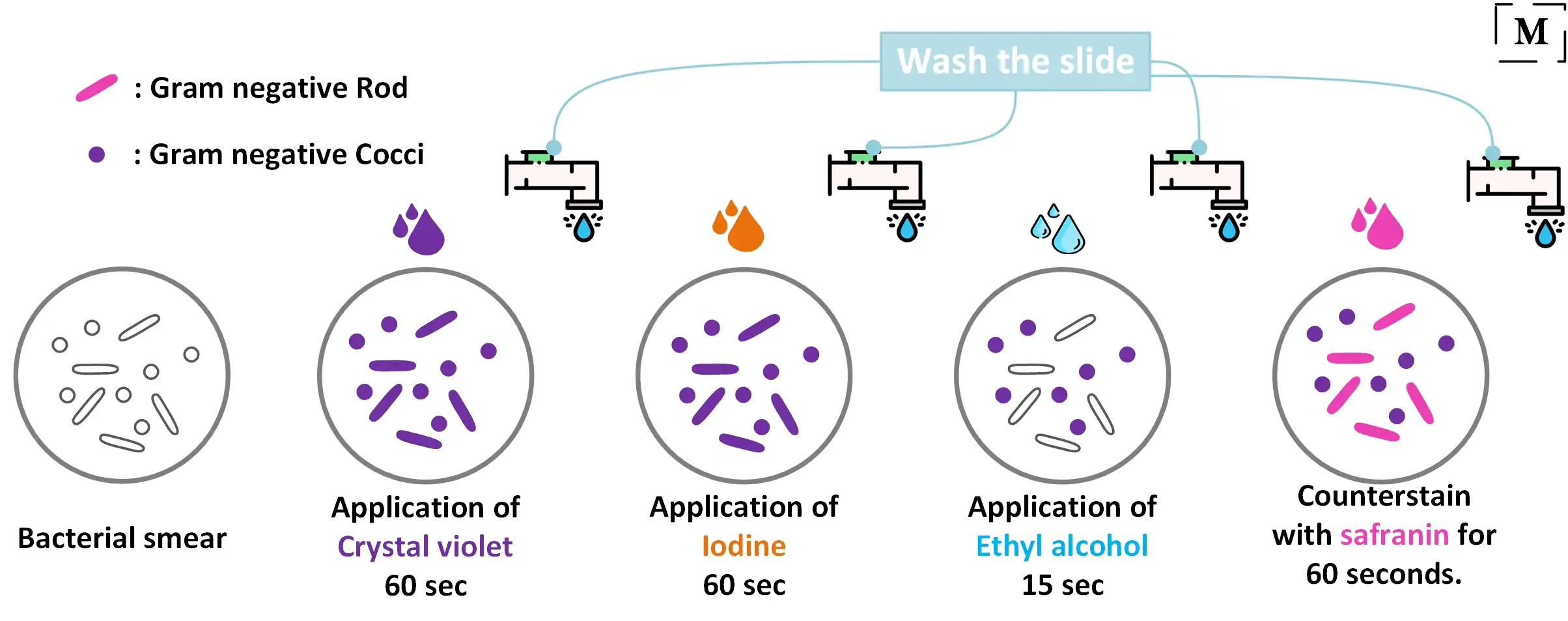
Decolorizing the cell causes this thick cell wall to dehydrate and shrink which closes the pores in the cell wall and prevents the stain from exiting the cell. Finally although not essential a counterstainof safraninis applied to the smear to dye the decolorized gram-negativecells with a pink color. If you dont proactively choose a different repayment plan option your federal student loans will default to the standard plan which has a term of.
Personal protective equipment 2. After decolorization step a counterstain is used to impart a pink. Gram-positive cells will stain purple and Gram-negative cells will stain red to pink.
Asked Mar 24 2017 in Biology Microbiology by NVdes. Both gram-positive and gram-negative cells have. The procedure is based on the ability of microorganisms to retain color of the stains used during the gram stain reaction.
Water tap water or. Gram Staining Principle. Bacteria cell walls are stained by the crystal violet.
The process of gram staining is used to differentiate between gram-positive and gram-negative bacteria notes. Supplies and Reagents 1. B The best use of a negative stain is.
To prevent the crystal violet from leaving the cells. Gram-positive cells will stain purple and Gram-negative cells will stain red to pink. Place the following steps in the correct sequence.
Gram staining targets the cell wall and a layer called peptidoglycan. Explain the difference between the constitutional and informal requirements to become president. You have performed a Gram stain and upon completion of the procedure the cells are colorless.
The next step also known as fixing the dye involves using iodine to form crystal violet- iodine complex to prevent easy removal of dye. The Gram stain involves staining bacteria fixing the color with a mordant decolorizing the cells and applying a counterstain. E The order doesnt matter.
Gram negative cells also take up crystal violet and the iodine forms a. The first step in gram staining is the use of crystal violet dye for the slides initial staining. The bacteria are differentiated through a series of staining and decolorization steps.
After the addition of the counterstain in gram staining the gram-positive bacterial cell appears purple whereas the gram-negative one appears pink. The primary stain crystal violet binds to peptidoglycan coloring cells purple. Place the following steps in the correct sequence to prepare the specimen.
View Test Prep - practical 1 study guide docx from BIOLOGY 101 at Pearl River Community College. A Gram stain is a test that checks for bacteria at the site of a suspected infection or in certain body fluids such as blood or urine. Iodine is subsequently added as a mordant to form the crystal violet-iodine complex so that the dye cannot be removed easily.
The Gram stain is important in medical microbiology because the results help physicians select appropriate antibiotics for treatment. Which step in the Gram stain is the critical step in differentiating gram-positive cells from gram-negative cells. To make gram-negative cells visible.
The purpose of a mordant in the Gram stain is. Since human cells do not have cell walls or peptidoglycan the gram stain would do nothing because the primary stain would wash out notes Wikipedia. Escucha la conversación entre daniel y.
Absorbent paper such as bibulous paper 5. Gram-negative bacteria are decolorized by the alcohol losing the color of the primary stain purple. Practical 1 study questions Cells are differentiated after which step in the Gram.
Bacteria having cell walls with a thick layer of peptidoglycan will resist decolorization of primary stain and appear violet or purple. The Gram stain procedure relies on structural differences of the cell walls between Gram-positive and Gram-negative bacteria that result in Gram-positive bacteria staining purple and Gram-negative bacteria staining pinkred. Cells are differentiated after which step in gram stain.
To make the bacterial cells larger. The procedure is based on the reaction between peptidoglycan in the cell walls of some bacteria. The organisms that do not take up primary stain appear red under a microscope and are Gram-negative organisms.
Cells are differentiated after which step in the Gram stain. This layer makes up 60-90 of the gram positive cell wall. The purpose of a mordant in the Gram stain is To remove the simple stain.
At the end of the gram staining procedure gram positive cells will be stained a purplish-blue color. The cell walls for Gram-positive microorganisms have a higher peptidoglycan and lower lipid content than gram-negative bacteria. Chemical and physical properties of their cell walls.
Gram Staining Better Understanding Of The Procedure And Easy Interpretation Of The Results
Why Does The Gram Positive Bacteria Stain Purple While Gram Negative Stains Pink Quora

Development Of A Standardized Gram Stain Procedure For Bacteria And Inflammatory Cells Using An Automated Staining Instrument Li 2020 Microbiologyopen Wiley Online Library


0 Comments